微奈米三維X-ray電腦斷層掃描儀

Xradia以超過全世界50個同步輻射加速器實驗室領域之實績, 在X-ray 3D斷層掃描影像(CT)分析產品被科學界及業界廣泛接受及使用.
目前, Xradia除在半導體封裝, 各式電路板產業(PCB)故障分析之成熟應用上,
X-ray 3D斷層掃描系列產品也已成功進入生物醫學,
生命科學, 石油和天然氣勘探,
奈米成像應用以及先進材料開發等相關領域.
Xradia是唯一提供在微奈米2D, 3D 及4D X-ray成像解決方案之設備供應商,
3D微奈米尺度之影像分辨率亦將引領您進入一個新的斷層掃描領域.

3D X-ray CT系列配有高可靠性, 免維護的封閉式X-ray射線源, 方便更換放大倍數的多透鏡偵測器及高穩定性的樣品台.
此產品亦可根據用戶設定而進行自動多點CT掃描成像,
實現24小時自動運轉之分析理想。
Micro/Nano-X-ray CT系列,
無需對樣品使用傳統的染色前處理,
對生物材料可快速進行非破壞性的表面和內部結構分析.
從半導體故障分析, 藥物開發, 分子影像,
幹細胞研究, 組織器官分析和先進材料的開發,
皆有相當成熟的應用。
- 非破壞性的3D成像
- 即時顯示成像過程
- 最少樣品製備需求
- 軟體指示多種倍率偵測器之選擇
- 自動連續操作多點斷層掃描
- 高解析度 (1μm ~ 50 nm)
- 分析樣品尺度大 (300mm)
Wirebond Shorts

BGA(BallGridArray) Defects

Battery Membrance
High Resolution Phase Contrast

Osteocyte Lacunar Properties
(骨細胞腔隙的性質)

Voids in Through-Silicon Vias (TSV)

應用解決方案
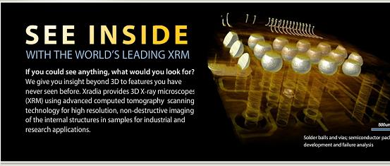
Semiconductor Process Optimization and Failure Analysis(半導體製程最佳化及失效分析)
Semiconductor packages are becoming more complex as the industry strives for ever increasing performance and efficiency. With this increased complexity, the associated failures are becoming much more difficult to determine and diagnose. Traditional methods such as physical cross sectioning and 2D X-ray imaging are no longer adequate. Today's failure analysis engineers need new techniques to assist them in their job. The Xradia VersaXRM high-resolution 3D X-ray microscope answers that need.
當這個工業永遠一直朝著增加性能及效率邁進, 半導體的組件就愈來愈複雜. 隨著複雜程度的增加, 連帶的失效問題也愈來愈難確認及診斷. 傳統的的方法如物理切斷面及二維X-Ray影像 不再夠用. 今日的失效分析工程需要新的技術來協助他們工作. Xradia 的 VersaXRM 高解析度三維X-Ray顯微鏡順應了這個須求.
With micron scale resolution available for a wide range of sample sizes and the ability to obtain high contrast on low Z materials, the VersaXRM is the premier 3D X-ray CT system available today. Maximum material and sample flexibility allow users to finally see defects in a truly non-destructive manner.
一個寬廣的樣品尺吋範圍內, 擁有微米尺度的解析度, 以及在低厚度材料可以得到高對比度的能力, VersaXRM 是今日可得到三維X-Ray電腦斷層掃描系統的首選. 最大的材料及樣品的彈性, 讓使用者終於能夠以真正的非破壞的方式看到缺陷的地方.
The VersaXRM gives the FA engineer the power of volumetric data. No more concern about cross sectioning exactly the right location. No longer will there be a question as to whether the inspection technique caused the defect or if the defect was already there. No more concern about ruining valuable defect samples. With the VersaXRM you can visualize a defect, non-destructively, and continue testing with the same package.
VersaXRM 給予工程失效工程師測量數據的力量. 再也不用顧慮必須精地地在正確位置作斷面切割的問題. 而諸如, 是否檢測技術本身有導致缺陷, 或則是本來就有否缺陷, 也不再是個問題. 不用再顧慮會損毀珍貴缺陷樣品. 用VersaXRM, 您可以非破壞地檢視一個缺陷, 並繼續樣品的測試.
Many difficult to diagnose FA problems become much easier to visualize and understand. Here are just a few of the many defects easily viewed with the VersaXRM:
許多難以診斷的失效分析問題變得容易觀察及理解多了. 這裏只是很多容易以VersaXRM來檢視的缺陷的一小部份:
Solder ball/bump voids and cracks(焊點球及接點間隙及裂痕).
Die attach voids and delamination(晶片接觸間隙及剝離)
Trace shorts and opens(微小的短路及開路)
Via cracks and opens(層板間通路的裂痕及開路)
Wirebond shorts and opens(銲線的短路及開路)
Solder reflow defects(焊接重熔的缺陷)
Life Sciences Solutions(生命科學的解決方案)
Xradia's VersaXRM high-resolution 3D X-ray microscope offers a complete range of powerful new capabilities, including: sub-micron pixel resolution 3D imaging, high contrast for imaging soft tissues and low atomic number materials, a unique system design that enables high resolution imaging of specimens in a variety of size, and shape alternatives. The system's large modifiable working distance also makes high resolution in-situ experimentation possible.
Xradia 的 VersaXRM 高解析度三維X-Ray顯微鏡, 提供了一個功能強大, 範圍完整的新能力, 包括: 次微米級解析度的三維影像, 對於柔軟組織及低原子數材料的高對比度, 能夠用於不同尺吋, 型狀檢體的高解析度影像的獨特的系統設計. 此系統大的可調的工作距離也使得高解析度的原位置實驗成為可能.
Because of its high resolution and high efficiency objectives, the VersaXRM has the ability to resolve features within biomedical samples like bone and tissue with great contrast. Xradia's unique utilization of magnification and optics makes the VersaXRM dominant within the X-ray imaging space in its abilities to image small and large samples with high resolution and contrast.
由於它的高解析度及高效率的物鏡, VersaXRM 具有以高對比度解析骨頭及組織樣品特徵的能力. Xradia 獨特的採用放大及光學設計, 使 VersaXRM, 以其用在小及大的樣品上的高解析度及對比度, 在X-ray影像的應用空間中取得了優勢.
The VersaXRM also addresses a wide range of biomedical research applications beyond histology such as:
High-resolution imaging of 3D vascular dispersion within organs, including angiogenesis mapping.
High resolution imaging of bone, calcified tissue, and cartilage samples to view features as small as osteocytes and trabecular bone density variations
Small animal imaging for evolution and growth studies, as well as teratology
Structural analysis of medical devices such as stents and catheters
除組織學外, VersaXRM也用到一個很大範圍的生物醫學的研究及應用, 例如:
器官內的血管分怖的高解析度三維影像, 包括血管生成的映像.
高解析度的骨骼, 鈣化的組織以及軟骨的樣品等的三維影像, 觀察小如骨細胞和小梁骨的密度變化.
小動物的演化及生長的研究的影像, 醫療器材的特異結構分析, 例如支架和導管.
Multi-Lengthscale Imaging Solutions
Visualizing and integrating the image data to describe a complex 3D biological organism or a functional material across length scales is challenging, especially since no single conventional imaging modality, such as visible, electron microscopy (EM) and AFM (atomic force microscopy) can cover the range. While EM and AFM can resolve structures to molecular and atomic scale, there is an ongoing problem in integrating these results into the larger scale structure and functions encountered in life science, geophysics or material science research. All imaging modalities require the right combination of field of view (FOV) and resolution to support navigation and orientation for region of interest (ROI) selection and high resolution and high contrast imaging.
要跨越一定長度尺吋, 視覺化及整合影像資料, 以描述一個複雜的三維有機體或是某種功用的材料, 是挑戰的, 特別是因為沒有傳統影像模式, 如, 可見光的, 電子顯微鏡, 原子力顯微鏡等, 能夠涵蓋這個範圍.
雖然電子顯微鏡及原子力顯微鏡能解析結構到分子及原子的尺度, 經常的有的問題是在於要把這些結果整合到在生命科學, 地質物理, 或材料科學研究的較大尺度及官能中. 所有的影像模式都需要正確的結合檢視區域及解析度, 以便能支援在感興趣區域的選擇及高解析度, 高對比度影像, 的目標要求時的導航與定向.
While 2D imaging modalities support "zoom in and out" for effective imaging, real non destructive 3D volume imaging does not support that capability. Xradia's multi length scale solution enables scientists and engineers to do this type of Scout-and-Zoom to effectively image samples of various scales at optimal resolution and field of view combinations.
雖然二維的影像模式支援有用的放大和縮小, 真實的非破壞三維量測影像並不支援這能力. Xradia 的多重長度尺吋解決方案讓科學家及工程師可以用這一類跟踪及變焦, 在最佳化的解析度及檢視區域的組合, 來有效地對不同尺度的樣品取得影像.
Encompassing the VersaXRM and UltraXRM platforms, Xradia's multi-length scale solution supports imaging from 40 mm FOV at ~30 micron resolution all the way down to 15 micron FOV at <50 nm resolution.
This supports complete 3D mapping of the internal characteristics of samples in a variety of research and engineering applications such as Hierarchical Framework Multiscale imaging in life science research, computational analysis in materials science studies, oil and gas drilling feasibility analysis and semiconductor package failure analysis.
圍繞於VersaXRM 及 UltraXRM的平台, Xradia 的多重長度尺吋解決方案, 支援從40mm的檢視區域(約30 micron解析度), 延著一直下到15mm的檢視區域(約50 micron解析度).
這支援了, 如在生命科學中的階層式架構, 在材料科學研究的計算分析, 的多重尺度的影像, 油氣鑽探的可行性分析, 以及半導體封裝失效分析, 等的各種研究及工程應用樣品的內部特性的完整三維影像.
Geo Materials Solutions
With the globally increasing demand for more energy, the world's oil and gas resources have been strained to its limits. As the price of oil increases, scientists are exploring every material available containing natural energy resources like oil and gas shale, carbonates, and limestone. Conventional core analysis techniques take months to produce viable results, causing production halts during analysis. The Xradia VersaXRM enables researchers to obtain pore connectivity and microstructure modeling on-site and within minutes.
隨著全球能源需求的增加, 全世界的石油及天然氣資源以緊繃於極限. 由於石油漲價, 科學家正探索每一種可取得的, 含有天然能量資源的材料, 如油及業岩, 碳酸鹽及石灰石. 傳統的礦核分析技術需要好幾個月以得出可行的結果, 在分析期間造成生產的停頓. Xradia VersaXRM 使研究人員能夠在幾分鐘之內, 就地得到孔隙連接程度及微結構的模式.
The VersaXRM can be adapted for mobility to enable on-site imaging of cuttings taken from core drilling.
Combining a high-flux laboratory X-ray source with proprietary X-ray optics into a compact ultra-high resolution CT scanner is useful for imaging large rock samples and also subsampling small cuttings for accurate extrapolation of large scale data.
VersaXRM 可以調整成可攜帶方式, 使其能夠就地對由礦核鑽探的切割物進行影像的取得.
結合一個高亮度的 X-ray 光源以及專有的X-ray光學設計到一個緊密的高解析度的電腦斷層掃描器, 對於大的岩石樣品的影像,
以及為了大尺吋數據的準確外插計算而做的小切塊的局部採樣, 是很有用的.
Because of its unique optics allowing for preferred detector and source setups, the VersaXRM can offer turnaround times on the order of minutes without compromising resolution or field of view.
Some of the applications the VersaXRM supports are:
Sandstone imaging for high-resolution grain orientation and pore connectivity
Limestone imaging for fossil detection and pore dispersion
Shale imaging for inclusions and micron sized pores
Oil and tar sands for high-resolution grain orientation, density dispersion, and pore connectivity.
由於它允許所比較喜歡的偵檢器及燈源的設置的獨特的光學設計, VersaXRM 能提供在幾分鐘程度的轉換時間, 而不需要妥協於解析度或檢視區域.
一些 VersaXRM 支援的應用包括:
用於高解析度的顆粒方向及孔隙連通性的砂岩影像.
用於化石偵測及孔隙散怖的石灰石影像.
油及焦油砂的高解析度顆粒方向, 密度散怖及孔隙連通性.
Materials Science
Xradia's VersaXRM provides an unparalleled combination of contrast and resolution for all types of materials science. From carbon fiber composite to high temperature ceramics to exotic metal alloys, the VersaXRM can meet all your 3D visualization needs.
With spatial resolution down to 1 micron for a variety of different sample types and sizes, the VersaXRM will provide true insight through 3D. The superior contrast of the VersaXRM makes it ideal for imaging low contrast, low Z materials. The volumetric data obtained from CT is ideal for applications from examining the effects of stress testing to identifying fiber orientations to measuring porosity.
在所有各種型式的材料科學方面, Xradia VersaXRM 提供了一個無與倫比的對比度及解析度的組合. 從碳纖維的組成到到高溫陶磁, 以及不尋常的金屬合金, VersaXRM能夠合於您的三維視覺需求.
對於各種樣品型式及尺吋, 可以小到1微米的解析度, VersaXRM將能夠提供真正的三維透視. VersaXRM優異的對比度, 使其可以理想地用於低對比度及低厚度的材料.
由電腦斷層掃描得到的量測數據, 在檢查應力試驗的效應, 以確認纖維的方向性, 量測其多孔性等應用是理想的.
With the expanded sample capacity of the MicroXCT-400, a member of the VersaXRM family, scientists and researchers have the added capability to perform in-situ testing of materials. The MicroXCT-400 can accommodate sample sizes up to 200 mm X 200 mm and a mass of up to 15 kg. The extra large enclosure can be used to house additional test equipment allowing for a large amount of flexibility for test set-up.
以VersaXRM家族的成員之一, MicroXCT-400的擴充的樣品容量, 科學家及研究人員已增加了進行材料就原位試驗的能力. MicroXCT-400 可適用的樣品尺吋達200 mm X 200 mm, 以及達15公斤的質量. 特大的容積可用於放入附加測試設備, 可允許很大的試驗設置的彈性.
Examples of materials commonly imaged using the VersaXRM:(常用VersaXRM
作影像的材料範例.
Fibers(纖維)
Composite Materials(複合材料)
Papers(紙)
Foams(泡沫)
Ceramics(陶磁)
Metals(金屬)
Thin Membranes(薄膜)
Woods(木材)
Concrete/Rocks(混凝土/岩石)
Fossils(化石)